

Blood Flow Restriction (BFR) Training: What it is and how it works
The use of BFR has increased in popularity over the past decade, particularly for its promising application in the clinical rehabilitation populations. And rightfully so! The body of literature supporting the use of BFR as an effective and safe tool is strong and growing.
 BFR was first introduced in Japan in 1966 and became popularized as “Kaatsu training”. It is now backed by decades of research. It was not until the early 2000s when tubes and wraps were replaced with “air cuffs”.
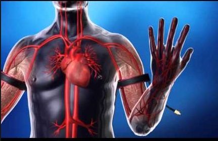
BFR utilizes pneumatic cuffs to allow arterial blood flow to a region while restricting venous return. BFR is not occlusion training. Occlusion training involves completely stopping blood flow into an extremity for significant periods of time which can damage tissue or nerves and increases the risk for medical conditions like DVTs and in extreme cases, rhabdomyolysis. Click on the images below to purchase these products!


The most common question when explaining BFR to those unfamiliar with the technique is: why would we want to do this?
 When venous return is limited, there is an accumulation of metabolic byproducts such as lactate. Lactate is needed as a buffering agent for the high concentration of hydrogen ions released during the hydrolysis of ATP, especially at higher absolute or perceived intensities of exercise.
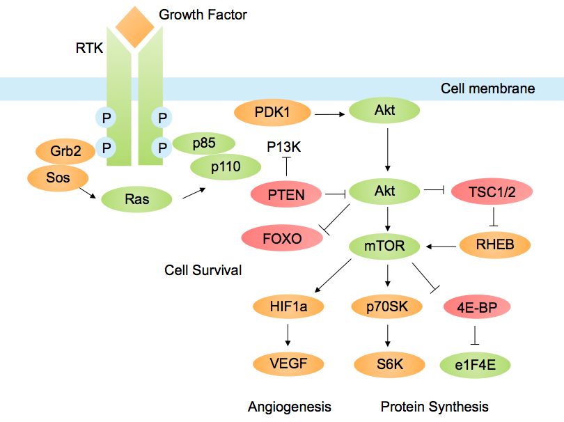
The accumulation of the metabolic byproducts leads to an increased acidic environment within the muscle. This disturbance of homeostasis begins a cascade of events that leads to an optimal anabolic environment. Increases in anabolic hormones like growth hormone and IGF-1 impact the signaling pathways that control protein synthesis (mTOR and mTORc1). Ultimately, this will lead to skeletal muscle hypertrophy over time.
In addition, BFR with exercise makes it more difficult to recruit Type I muscle fibers and the threshold for recruiting Type II fibers (our main strength and power muscle fibers) is lowered. This leads to a recruitment pattern opposite of the traditional “size principle of fiber recruitment.”
Typically, the above beneficial anabolic environment and physiological adaptations are only achieved during high intensity and high volume strength training. But associated with this type of training is the risk of muscle breakdown, joint stress, and non-contractile tissues.
In future blog posts, I will discuss the safety of BFR, common myths, and why the use of BFR in addition to low intensity resistance exercise in the clinical populations is a great tool for clinicians.

Make sure to check out the Modern Strength Training: Blood Flow Restriction live seminar being held August 5-6, 2017 - register here!
Feel free to contact me with any questions!
Website: www.motusptperformance.com
Facebook: www.facebook.com/motusptperformance
Instagram: @motuspt
Email: Kyle.MotusPT@gmail.com

Keeping it Eclectic...

 |
| Learn More |
Want an approach that enhances your existing evaluation and treatment? No commercial model gives you THE answer. You need an approach that blends the modern with the old school. Live cases, webinars, lectures, Q&A, hundreds of techniques and more! Check out Modern Manual Therapy!
Keeping it Eclectic...

References
- Sato Y. The History and Future of Kaatsu Training. Int J. Kaatsu Training Res. 2005; 1: 1-5.
- Manini TM, et al. Blood Flow Restricted Exercise and Skeletal Muscle Health. Exerc. Sport Sci. Rev. 2009. 37(2): 78-85.
- Manini TM, et al. Blood Flow Restricted Exercise and Skeletal Muscle Health. Exerc. Sport Sci. Rev. 2009. 37(2): 78-85.
- Kawada S, et al. Changes in skeletal muscle size, fibre-type composition and capillary supply after chronic venous occlusion in rats. Acta Physiol. 2008. 192: 541–549
- Slysz J, et al. The efficacy of blood flow restricted exercise: A systematic review & meta-analysis. Journal of Science & Medicine in Sport. 2016; 19: 669-675.
- Loenneke JP, et al. Low intensity blood flow restriction training: a meta-analysis. European Journal of Applied Physiology. 2012. 112(5): 1849-1859.
- Soto GA, et al. The Effects of Short-Term Resistance Training with & without Blood Flow Restriction on Neuromuscular Adaptations. International Journal of Exercise Science. 2017. 2(9): Article 14.
- Karabulut M, et al. Overview of neuromuscular adaptations of skeletal muscle to KAATSU Training. International Journal of Kaatsu Training Research. 2007. 3: 1-9.
- Yasuda T, et al. Muscle fiber cross-sectional area is increased after two weeks of twice daily KAATSU-resistance training. Int. J. KAATSU Training Res. 2005. 1: 65-70.
- Takarada Y, et al. Cooperative Effects of Exercise and Occlusive Stimuli on Muscular Function in Low-Intensity Resistance Exercise with Moderate Vascular Occlusion. Japanese Journal of Physiology. 2004. 54: 585–592.
- Abe T, et al. Muscle Size and IGF-1 Increased after Two Weeks of Low-Intensity “Kaatsu” Resistance Training. Medicine & Science in Sports & Exercise. 2004. 36(5) Supplement.
- Madarame H, et al. Endocrine responses to upper- and lower-limb resistance exercises with blood flow restriction. Acta Physiologica Hungarica. 2010. 97(2): 192–200.
- Gundermann DM, et al. Reactive hyperemia is not responsible for stimulating muscle protein synthesis following blood flow restriction exercise. Journal of Applied Physiology. 2012. 112: 1520-1528.


 Training: What it is and how it works The use of BFR has increased in popularity over the past dec...)
Post a Comment
Post a Comment